BIOMIMESYS® Oncology
3D cell culture model for tumor cells
Product Basics
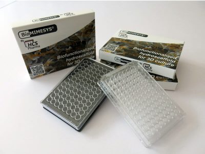
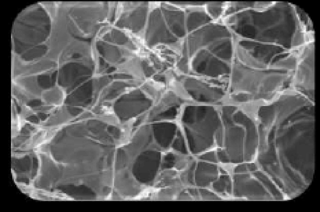
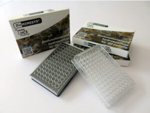
BIOMIMESYS® Oncology is a Hyaluronic acid (HA) based scaffold 3D cell culture model. This groundbreaking 3D cell culture technology associates the behavior of a solid scaffold and of a hydrogel, which we call “Hydroscaffold”. The Hydroscaffold’s composition and mechanical properties can mimics the microenvironment of tumor cells. This scaffold provided cell binding sites with CD44 and RHAMM allowing cell-matrix and cell-cell interactions. Also, proven to provide better in vitro/in vivo correlation than 2D models.
Key Features
- Recreate the mechanical stress (stiffness) of the tumor cell’s ECM
- Better in vitro/in vivo correlation
- Provides cell binding sites with CD44 and RHAMM
- Ready to use
- Applicable for most downstream applications
- Batch to Batch consistent
Technical Information
Better reproducibility of mechanical stress on cells
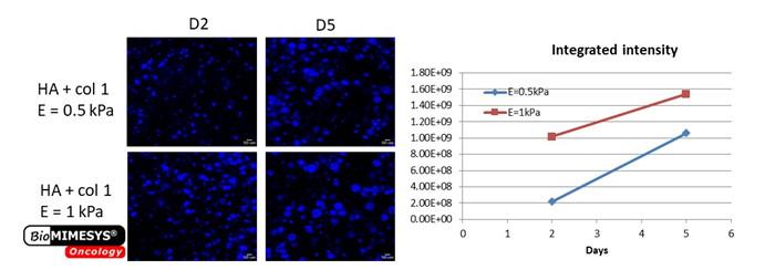
Mechanical stress in solid tumors can impede drug delivery and may additionally drive tumor progression and promote metastasis. Mechanistically, biomechanical forces can drive tumor aggression by inducing a mesenchymal-like switch in transformed cells so that they attain tumor-initiating or stem-like cell properties. ¹ (Northey et al, 2017)
Above shows the increase in cellular proliferation in response to mechanical stress (2x in stiffness) of 3D matrices. Mechanical stress on cells can be mimicked by increasing in solid fiber (GAGs and collagens) concentration with BIOMIMESYS ®. This cannot be reproduced on 2D or 3D cell culture using hydrogels or Matrigel.
Better in vitro/in vivo correlation
By using BIOMIMESYS® Oncology, anti-oncogenic drugs (such as 5-Fluorouracil, 5-FU) showed cytotoxic effects closer to human responses compared to 2D culture.
Ready to use
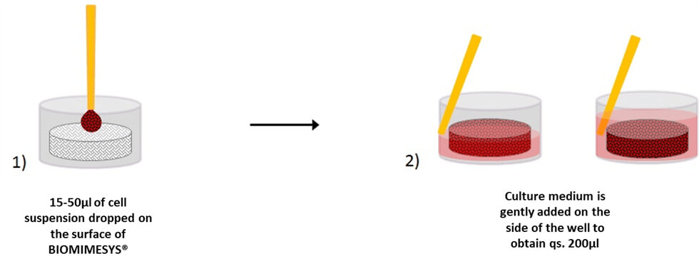
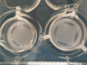
BIOMIMESYS® Oncology is EASY & READY TO USE. Upon receiving the vacuum sealed 96-well plate open it (under a hood) and add the cells directly on top of the matrix. Changing the culture medium is easy as well. To remove medium, simply draw the medium with a pipette between the matrix and the edge of the well. To refresh the medium, place fresh medium onto the surface of the matrix.
Successfully Tested Cells
Cell Lines
Human brain metastasis: SA87
Human breast adenocarcinoma : MCF-7
Human breast carcinoma : CAL-51
Human cervix adenocarcinoma : HeLa
Human colorectal adenocarcinoma : DLD-1, HT29, Caco-2
Human glioblastoma : CB109 / CB74 / CB191
Human liver hepatocellular carcinoma : HepG2
Human liver hepatoma : PLC / PRF-5
Human lung carcinoma : NCI-H460
Human osteosarcoma : SaOs
Human ovarian carcinoma : IGROV-1
Human pancreas carcinoma : PANC-1
Human prostate cancer : PC3
Normal human colon fibroblast : CCD18-co
Stem Cells
Hematopoietic : CD34+
Human induced pluripotent stem cells : hiPS
Complete List of Tested Cells [PDF]
Compatible with Most Downstream Applications
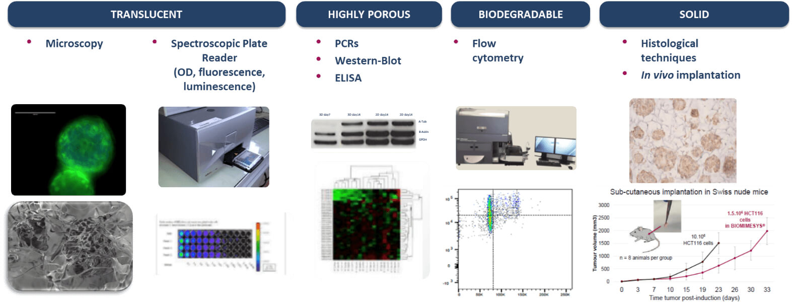
BIOMIMESYS® hydroscaffold has many properties (transparent, porous, biodegradable, & solid) making it ideal for use with numerous downstream applications.
Specification
- Hyaluronic acid (HA) (1.6MDa) – Provides binding sites for CD44 and RHAMM cell surface receptors
- Adipic acid dihydrazide (ADH) as crosslinker
- Collagen Type I
- Young’s modulus: 1.1kPa – different stiffness is available by customization
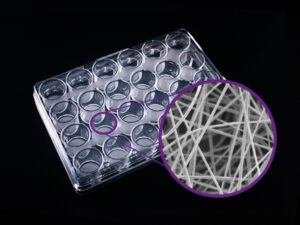
- Porosity: 155±35µm
Pricing
24 Hydrogel / 96 Well
Transparent Plate
- SKU: BIO_ONC_96_24_transp
- Price:
$410.00→ $369.00
Black Plate
- SKU: BIO_ONC_96_24_black
- Price:
$410.00→ $369.00
96 Hydrogel / 96 Well
Transparent Plate
- SKU: BIO_ONC_96_96_transp
- Price:
$590.00→ $531.00
Black Plate
- SKU: BIO_ONC_96_96_black
- Price:
$590.00→ $531.00
384 Hydrogel / 384 Well / Black
1 Plate
- SKU: BIO_ONC_384_384_black
- Price:
$665.00→ $600.00
5 Plates
- SKU: BIO_ONC_384_384_black_5
- Price:
$2500.00→ $2250.00
References & Literature
1. Bubba, F. et al. A chemotaxis-based explanation of spheroid formation in 3D cultures of breast cancer cells. Journal of Theoretical Biology 479, 73–80 (2019) doi: 10.1016/j.jtbi.2019.07.002.
2. Lane, R. et al. Cell-derived extracellular vesicles can be used as a biomarker reservoir for glioblastoma tumor subtyping. Communications Biology 2, 315 (2019) doi: 10.1038/s42003-019-0560-x.
3. Gomes, A. et al. Evaluation by quantitative image analysis of anticancer drug activity on multicellular spheroids grown in 3D matrices. Oncol Lett 12, 4371–4376 (2016) doi: 10.3892/ol.2016.5221.
4. Salaroglio, I. C. et al. Increasing intratumor C/EBP-β LIP and nitric oxide levels overcome resistance to doxorubicin in triple negative breast cancer. Journal of Experimental & Clinical Cancer Research 37, 286 (2018) doi: 10.1186/s13046-018-0967-0.
5. Vitali, E. et al. Metformin and Everolimus: A Promising Combination for Neuroendocrine Tumors Treatment. Cancers 12, (2020) doi: 10.3390/cancers12082143.
6. Salaroglio, I. C. et al. PERK induces resistance to cell death elicited by endoplasmic reticulum stress and chemotherapy. Molecular Cancer 16, 91 (2017) doi: 10.1186/s12943-017-0657-0.
7. Simon, T. et al. Shedding of bevacizumab in tumour cells-derived extracellular vesicles as a new therapeutic escape mechanism in glioblastoma. Molecular Cancer 17, 132 (2018) doi: 10.1186/s12943-018-0878-x.
8. Northey, J. J., Przybyla, L. & Weaver, V. M. Tissue Force Programs Cell Fate and Tumor Aggression. Cancer Discov 7, 1224 (2017) doi: 10.1158/2159-8290.CD-16-0733.
Other Documents
- Retrieve Cells / Washing Gels
- First Steps - Co-culture, Change Medium, Seeding
- Protocol for Metabolic Activity Determination
- RNA or DNA Extraction
- Protein Extraction
- Microscopy
- Flow Cytometry
- Preparation of Samples for Histological Labeling
- Evaluation of BIOMIMESYS® hydroscaffold in mice bearing subcutaneous HCT 116 Human Colorectal tumour cells
- Technical Datasheet BIOMIMESYS® Oncology
- MSDS BIOMIMESYS® Oncology
FOR RESEARCH USE ONLY, NOT FOR USE IN DIAGNOSTIC PROCEDURES
